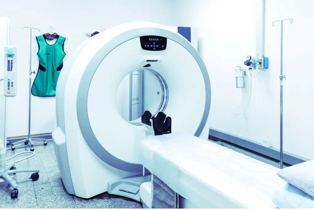
CT Scans in Diagnosing Mesothelioma are a pivotal tool for medical professionals. Mesothelioma is a rare but aggressive cancer that primarily affects the lining of the lungs, abdomen, or heart. The most common type of mesothelioma is pleural mesothelioma, which occurs in the pleura—the lining of the lungs.
The disease is primarily associated with a history of asbestos exposure, often affecting asbestos-exposed workers. The diagnosis of mesothelioma involves multiple tests, with CT (Computed Tomography) scans playing an essential role.
Mesothelioma and Asbestos
Asbestos exposure is the primary cause of mesothelioma, a type of cancer that affects the lining of the lungs and abdomen. When asbestos fibers are inhaled, they can become embedded in the soft tissues of the body. It can lead to the formation of pleural plaques, pleural thickening, and eventually, malignant mesothelioma.
Moreover, The link between previous asbestos exposure and the development of mesothelioma is well-documented and widely accepted in the medical community.
However, it often takes a clinical latency period of 35-40 years from the time of exposure for symptoms to appear. Moreover, making it a particularly insidious disease. This prolonged latency period often complicates the diagnosis and treatment of mesothelioma.
Pleural Changes and Mesothelioma
In the early stages of mesothelioma development, the disease may cause a variety of pleural abnormalities. It include pleural effusions (fluid in the pleural space), pleural thickening, and pleural plaques. These changes in the pleura make it difficult for physicians to arrive at a differential diagnosis as they can also be associated with other diseases.
Therefore, advanced imaging tests such as CT scans, PET scans, and magnetic resonance imaging (MRI) become crucial in determining the extent of the disease and making an accurate diagnosis. These imaging techniques can provide detailed images of the pleura and help identify any abnormal growths or changes. It is essential for planning the appropriate treatment options.
CT Scans in Diagnosing Mesothelioma
CT scans, or computed tomography scans, are instrumental in the diagnosis of mesothelioma. They produce detailed cross-sectional images of internal structures, making them ideal for observing abnormalities like pleural rinds, tumor nodules, and changes in the pleural surface. CT scans are often considered the gold standard for initial imaging. Because they offer greater detail than standard X-rays or chest radiography.
Pleural Mesothelioma
In cases of pleural mesothelioma, which affects the lining of the lungs, CT scans can detect pleural effusions (fluid in the pleural space), pleural thickening, and even mediastinal shift (movement of the central chest structures). These findings are crucial for making an accurate diagnosis and determining the extent of the disease.
Peritoneal Mesothelioma
Peritoneal mesothelioma affects the lining of the abdomen and can cause peritoneal thickening and the formation of tumor nodules. CT imaging can help in these cases by showing cross-sectional images of the peritoneal surfaces, aiding in the definitive diagnosis. This is especially important in peritoneal mesothelioma as it may not present with symptoms until advanced stages.
Advanced Imaging Techniques
PET Scans
Positron emission tomography (PET) scans are essential tools in assessing the metabolic activity of cells. When combined with CT scans, as in 18F-FDG PET/CT, it offers a 3-D image of the tumor. It reveals not just its structure, but also its activity. This combination is crucial for accurately staging the cancer and understanding its extent, which in turn helps in planning the treatment strategy.
MRI
Magnetic resonance imaging (MRI) utilizes magnetic fields and radio waves to generate detailed images of the body’s internal structures. This makes it an invaluable tool for resolving soft tissue structures and can further validate CT findings, especially when there is pleural or peritoneal involvement. It is often used in conjunction with other imaging techniques to provide a comprehensive view of the tumor and its impact on surrounding structures.
Needle Biopsy and Tissue Samples
After the imaging tests, a needle biopsy often follows to obtain tissue samples for testing. This step is crucial for a definitive diagnosis of mesothelioma. Surgical biopsies can also be performed, depending on the clinical presentation.
Staging and Treatment Options
The stage of mesothelioma is a crucial aspect of managing the disease and is typically determined through CT scans, PET scans, and other diagnostic methods like biopsy. The stage indicates the extent of disease spread, whether it is localized or has metastasized to other parts of the body. Advanced disease stages may show tumor growth into the chest wall, direct extension into other organs, or even metastatic carcinoma. The treatment options for mesothelioma are largely dependent on the stage of the disease. Early stages can manage with surgical treatment to remove the tumor.
However, in advanced stages where the tumor has spread extensively, systemic treatment like chemotherapy may be the preferred option. you can consider other treatment options such as radiation therapy, depending on the extent of the disease and the overall health status of the patient. Ultimately, the choice of treatment is individualized, taking into account various factors including the stage of mesothelioma, the patient’s overall health, and the potential benefits and risks of each treatment option.

FAQs:
What does mesothelioma look like on a CT scan?
Mesothelioma can appear on a CT scan as a thickening of the pleura, pleural effusions, and tumor nodules. The CT images can also show any mediastinal shift or involvement of other organs or tissues.
Can a CT scan show mesothelioma?
Yes, a CT scan is often considered the gold standard for initial imaging in mesothelioma diagnosis.
Can mesothelioma be missed on a nuclear CT scan?
While CT scans are highly effective in detecting mesothelioma and its associated changes, no imaging test is 100% foolproof. There is always a small chance that the disease could be missed, especially in its early stage. A nuclear CT scan, which combines a CT scan with a radioactive tracer. It can provide more detailed information about metabolic activity and may increase the chances of detection.
Does mesothelioma show up on a CT scan?
Yes, mesothelioma typically shows up on a CT scan as pleural thickening, pleural effusions, and tumor nodules on the pleural surface. However, the appearance of mesothelioma can vary from person to person and may be influenced by the stage of the disease.
What are the signs on X-ray or CT scan of mesothelioma?
Signs of mesothelioma on an X-ray or CT scan may include pleural thickening, pleural effusions, and tumor nodules on the pleural surface. On a CT scan, you may also see changes in the mediastinal structures and any involvement of other organs or tissues. An X-ray may show less detail but can still be useful in identifying pleural effusions and significant pleural thickening.
Conclusion
CT scans in diagnosing mesothelioma is very important. It is especially valuable for detecting pleural effusions and pleural diseases, which are often the first indicators of mesothelioma in patients with a history of asbestos exposure. When combined with other imaging methods like PET scans and MRI, as well as tissue samples obtained through biopsy, CT scans contribute significantly to the accurate diagnosis and treatment planning for mesothelioma patients.

