
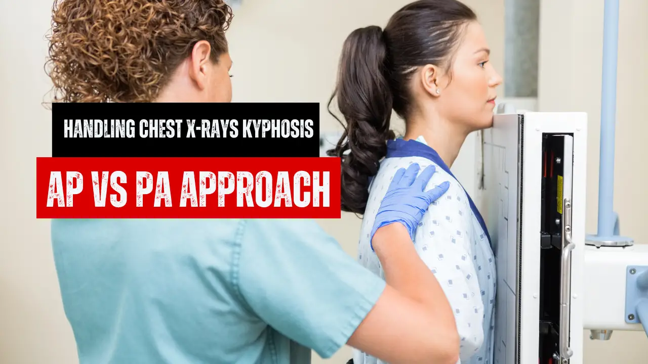
When it comes to ‘Handling Chest X-rays Kyphosis’, the radiology sector often finds itself at crossroads.
This article presents an interesting case of a patient with severe kyphosis and our approach to their radiographic imaging.
Kyphosis Severity and the Chin Conundrum
We must first consider how severe kyphosis is to unravel this dilemma. In extreme kyphosis cases, the PA approach gives an OID from the heart equivalent to or more significant than what we’d get from an AP approach.
We need to get the patient’s chin out of the way to nail the PA. A PA could be your best bet if the patient can manage that. However, AP might be your friend if the chin remains an obstacle.
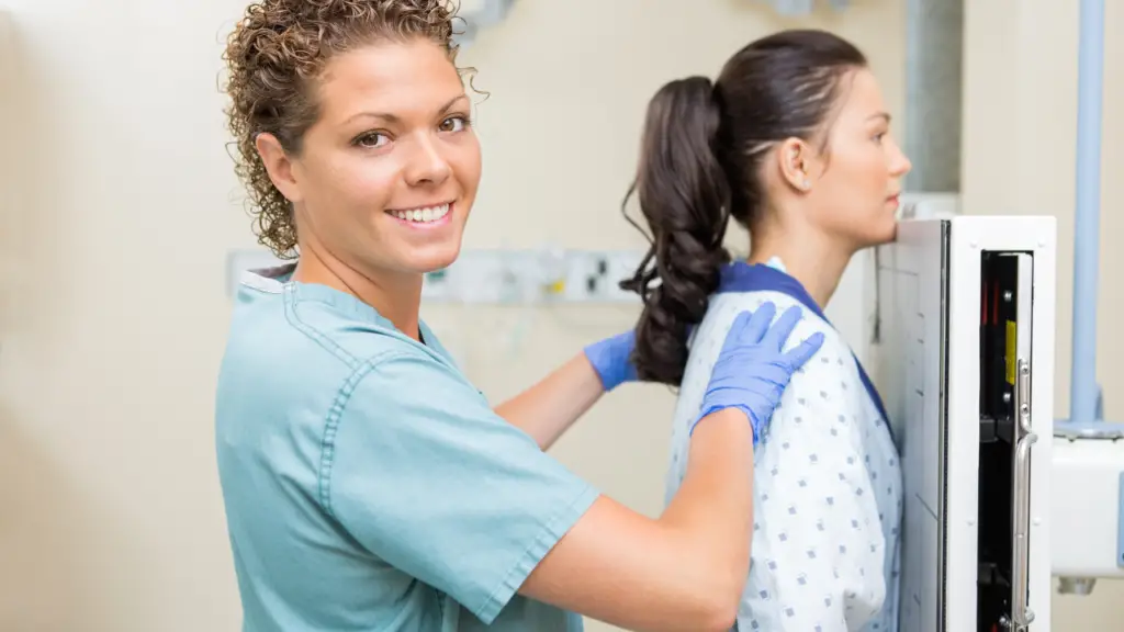
In my case, the patient was significantly kyphotic and couldn’t lift their chin enough. So, AP was the day’s hero, bringing the patient closer to the Image Receptor (IR).
The Handy Stool Technique
Here’s a fun tip that might come in handy. Consider bringing a stool into the picture. A seat can help the patient stand straighter and get closer to the wall Bucky, making a PA projection with a caudal angle easier to achieve.
Seasoned Techs’ Quick Solution
But let’s not complicate things unnecessarily. Some seasoned techs swear by the old-school ‘hug the Bucky’ PA method, widen the collimation and get the job done without fuss.
The X-Ray Tube Angle Game
Playing around with the angle of the X-ray tube saves the day. In my case, I took a straight-on PA shot first, followed it up with another image using a caudal angle, raising the tube quite a bit for this second shot.
The Doctor’s Wish is Our Command
Remember, what we’re after might differ depending on what the doctor is searching for. We should focus on getting a clear image if they need a good look at the heart. If lung issues or masses are the concern, we must strive to capture the entire lungs in all their glory.
AP Approach to the Rescue
With extreme kyphosis patients, the less conventional AP approach could sometimes be the real game-changer. It can help us get a diagnostic image and open up the lung apices while keeping the beam horizontal for those pesky air/fluid levels.
FAQs
What is the usual approach for handling chest X-rays in severe kyphosis cases?
The AP approach is typically used; however, if the patient can get their chin out of the way, the PA approach could be more effective.
How does the severity of kyphosis affect the radiologic approach?
In cases of extreme kyphosis, the patient’s posture could increase the OID from the heart, affecting the clarity of the image. In these instances, altering the approach may be necessary.
Can the AP approach be useful in handling chest X-rays for severe kyphosis?
Yes, especially when patients cannot lift their chin enough for a clear PA image. The AP approach can bring the patient closer to the Image Receptor (IR), thus yielding a more diagnostic image.
Conclusion
Handling Chest X-rays Kyphosis” can indeed present a challenge, but it’s through these challenges that we learn and grow as practitioners.
By tailoring our approach based on the patient, the specific clinical scenario, and the radiologist’s preferences, we can aim for the best diagnostic image with maximum patient comfort.
Let’s continue to ask questions, learn, and enhance our daily radiology practice.
