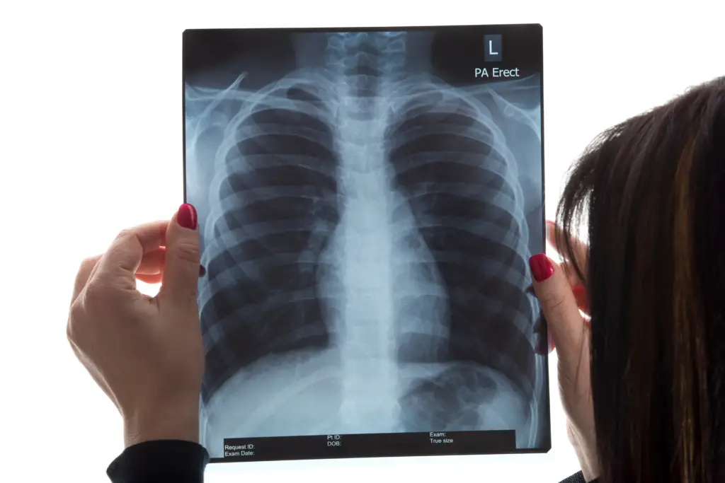
A chest radiograph, commonly known as a chest X-ray or CXR, is often reputed to be the most widely conducted radiological investigation globally. However, this widely held belief doesn’t have any published data for validation.
Still, data from the NHS in England and Wales suggest that the essentials of chest radiography remain the top requested imaging test by GPs, according to the 2019 dataset.
For pediatric specifics, refer to the chest radiograph (pediatric) section.
Indications
A chest radiograph is vital for an array of medical indications, including:
- Respiratory and cardiac diseases
- Hemoptysis and suspected pulmonary embolism
- Investigations into tuberculosis, pneumonia, and pneumothorax
- Suspected metastasis
- Monitoring disease progress and chronic dyspnea
- Trauma evaluations
- Checking positions of nasogastric tubes, endotracheal tubes, etc.
- Excluding radiopaque foreign bodies, such as for MRI safety checks
- And many more, including pre-employment screenings and immigration checks.
Projections
1. PA Projection:
- Standard frontal chest projection, where the x-ray beam goes from posterior to anterior.
- Advantages: Excellent visualization of the mediastinum and lungs.
- Disadvantage: Requires the patient to stand.
2. AP Projection:
- An alternative to the PA, the beam travels from anterior to posterior.
- Advantages: Suitable for patients who can’t stand.
- Disadvantages: Mediastinal structures may seem magnified; the projection is often rotated and may create skin folds.
3. Lateral Projection:
- Performed erect left lateral, it helps in locating suspected chest pathology.
- Note: The radiation dose from this is notably higher than a PA projection.
4. Additional Projections: These include:
- Lateral decubitus
- Expiration view
- Lordotic view
- RAO/LAO view
- Ribs AP/PA view
- Sternum lateral/oblique view
Common Pitfalls
- Rotations can drastically change the CXR appearance.
- Supine positioning can alter the appearance, enlarging the heart and changing fluid or gas appearances.
- Skin folds might resemble signs of pneumothorax.

Patient Preparation
Before undergoing a chest radiograph:
- Patients should remove upper body clothing and jewelry.
- Hair should be tied up.
- Tubings and lines must be out of the radiography field.
Dosage Information
The voltage used during the procedure affects the resulting image and patient exposure. High kV (120-140 kV) is more efficient than low kV (60 – 70 kV) in showcasing the lungs behind major organs. Plus, the higher kV helps reduce the patient’s effective dose.
To give you a sense, the effective dose of a chest X-ray (combining PA and lateral views) varies between 0.06 to 0.25 mSv, depending on the system and voltage used. The PA view takes up 25% of the effective dose, while the lateral view accounts for the remaining 75%.

