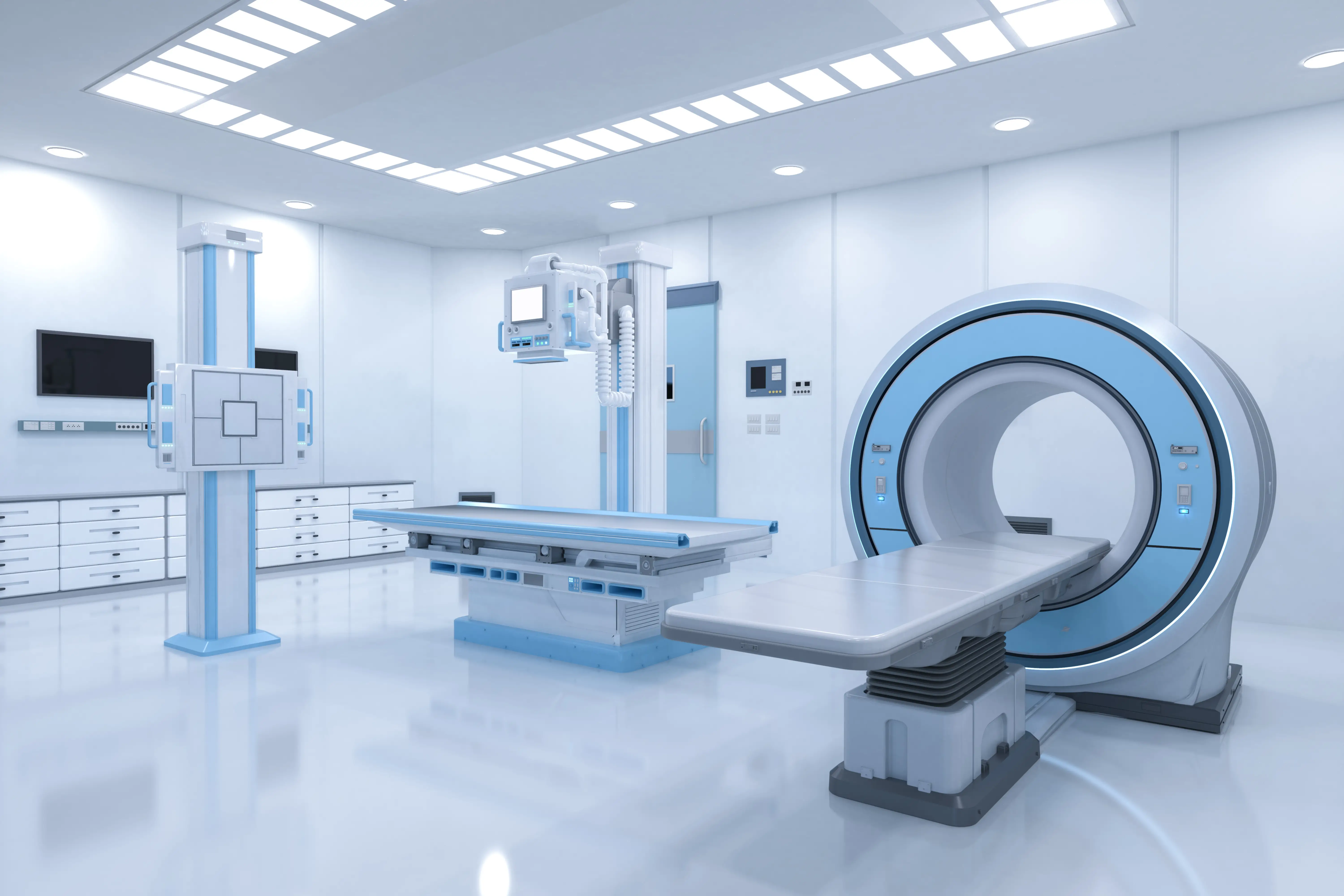
In medical science, cardiac imaging stands as a pivotal element in diagnosing and managing heart diseases. This sophisticated field has revolutionized our approach to heart-related ailments, particularly in conditions like coronary artery disease and valvular heart diseases. Moreover, cardiac imaging plays a crucial role in guiding interventional procedures, ensuring precision and enhancing patient outcomes.
Coronary Artery Disease through Imaging
Coronary artery disease (CAD), a leading cause of mortality globally, necessitates early and accurate detection. Advanced imaging techniques have become instrumental in diagnosing CAD. Techniques such as Coronary Computed Tomography Angiography (CCTA) offer a non-invasive yet highly detailed view of the coronary arteries, detecting blockages and plaque buildup. Additionally, Myocardial Perfusion Imaging (MPI) using Single Photon Emission Computed Tomography (SPECT) or Positron Emission Tomography (PET) provides insights into the blood flow to the heart muscle, crucial for assessing the severity of CAD.
Valvular Heart Diseases
Valvular heart diseases, which involve the dysfunction of one or more of the heart valves, demand precise imaging for accurate diagnosis. Echocardiography remains the cornerstone of valvular disease evaluation, offering real-time images of valve motion, and assessing the severity of valve stenosis or regurgitation. Advanced techniques like Transesophageal Echocardiography (TEE) provide even more detailed views, especially in cases where transthoracic images are not sufficient.
The Evolution of Cardiac MRI
Cardiac Magnetic Resonance Imaging (MRI) has emerged as a game-changer in the field of cardiac imaging. Its ability to provide high-resolution images of the heart’s structure and function without radiation exposure is unparalleled. It is particularly useful in assessing complex congenital heart diseases, cardiomyopathies, and in the evaluation of myocardial viability post-myocardial infarction.

Cardiac CT
Cardiac Computed Tomography (CT) has evolved significantly, now providing high-resolution, three-dimensional images of the heart and vessels. This technique is particularly beneficial in evaluating coronary artery anomalies, aortic diseases, and in pre-procedural planning for various cardiac interventions.
Nuclear Cardiology
Nuclear cardiology, utilizing techniques like SPECT and PET, plays a critical role in the functional assessment of the heart. These methods are essential in evaluating myocardial perfusion, ventricular function, and in identifying viable myocardium in patients with ischemic heart disease.
The Role of Imaging in Guiding Interventional Cardiology Procedures
Cardiac imaging is not just diagnostic; it is integral in guiding interventional cardiology procedures. Techniques like Intravascular Ultrasound (IVUS) and Optical Coherence Tomography (OCT) provide detailed images from inside the coronary arteries, guiding stent placement and ensuring optimal outcomes. Fluoroscopy remains a mainstay in guiding catheter-based interventions.
Future Directions in Cardiac Imaging
The future of cardiac imaging is bright, with ongoing advancements aimed at enhancing resolution, reducing exposure to radiation, and increasing the speed of image acquisition. Artificial Intelligence (AI) and Machine Learning (ML) are set to play a significant role in image interpretation, potentially improving diagnostic accuracy and patient care.
Conclusion
Cardiac imaging has become indispensable in modern medicine, providing a window into the intricate workings of the heart. Its role in diagnosing and managing heart diseases, particularly coronary artery disease and valvular heart diseases, is invaluable. As technology advances, we can anticipate even greater strides in this crucial field, further enhancing patient care and treatment outcomes.

