
Radiology protocols can vary depending on a few things:
- Radiologist preferences
- Hospital preferences
- Limiting factors based on equipment
But for the most part, radiology protocols are very similar to some degree. Today, I’m sharing a basic Barium Enema protocol just in case someone needs it for their clinic.
Barium Enema Protocol
Supplies
- IV Pole
- KY Jelly
- Blue Bulb Air Pump (whatever pump you use)
- Balloon Inflator
- Double XL Enema System (enema kit)
- Liquid Polibar
- Green Clamps (clamps to close off tubing)
- Chucks (cloth absorbant pad)
- Tape
- Glucagon available if Radiologist wants to administer it
Barium Enema Setup & Procedure
- Set up the room based on your Radiologist’s preferences.
- Perform a Timeout with Radiologist (right patient, right procedure, right site)
- Explain the procedure to the patient.
- Obtain a Scout KUB image.
- Show the scout image to the Radiologist and the exam order.
- Move patient to their left side and tip (if you are allowed)
- Radiologist will tell you whether or not to do post images
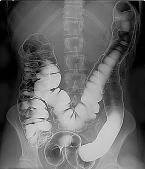
- Capture the following images:
- AP angled sigmoid
- AP KUB
- Both Decubitus images
- PA KUB
- Cross-table lateral rectum with tip OUT
- Post evac KUB
Post Procedure
- Do not let the patient leave until you show images to the Radiologist.
- A radiologist determines if the patient gets to leave.
Show the images to the Radiologist before letting the patient leave. Follow Radiologist guidance on any additional images.
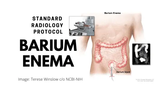
What is a Barium Enema – Tech Refresher
A barium enema is another term for an xray of the colon. Also called a B.E., it can reveal abnormal pathology in the large intestine (colon).
The enema is an injection of liquid into your rectum through a small tube. The liquid is a barium contrast that shows up on imaging.
The barium will outline the colon walls and show the gross characteristics of the colon.
