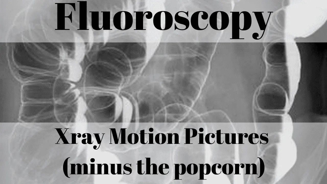
What is a fluoroscopy scan?
- a real-time image capture in motion picture format of human anatomy using continuous xray beam technology
As a technologist, I have used fluoroscopy in just about every fashion of its intended use. From pain injections in outpatient clinics to hip replacement surgeries in hospital operating rooms. What amazes me about this piece of imaging technology is how mobile and versatile it can be and still deliver superior diagnostic quality images. Nothing else provides real-time xray imagery like a fluoroscopic piece of equipment. For this article, I won’t touch much on endoscopic uses as radiologic technologists typically do not participate in those studies. They are done by the physician. Technologists operate equipment for fluoroscopy uses typically in the general imaging department, interventional procedures, and the operating room.
Fluoroscopy is an imaging instrument used for observing the internal structure of an object. In the field of medical diagnosis, it used for imaging the internal structure of the living body. An X-ray beam passes through the body; the beam then transmits to a TV-like screen. The screen enables the operator to examine the body in detail. It displays the real-time image for better diagnosis results. The fluoroscopy procedure list includes exploring the flow of blood for arterial blockages, the movement of intestines, abnormal connection between organs, and more.
A fluoroscopy procedure is also helpful for treating compression fractures of the spine and guiding injections into joints or the spine. A fluoroscopic injection is used to identify the source of the pain or assure that the pain is bearable. It is an excellent source of pain management treatment. The fluoroscopic indications are useful in various orthopedic procedures, gastrointestinal investigations, cardiovascular and interventional radiology procedures. It may assist in the insertion of implants in cases of fractured bones and position the alignment. Fluoroscopy can also help to monitor progress in matters of catheter insertion.

How do I prepare for fluoroscopy?
A fluoroscopy procedure may vary according to the condition of the person. There are, however, some special preparations before the process. Some of the generally followed fluoroscopy procedure preparations mentioned below:
- Some fluoroscopic procedures require patients to be in a fasting state for the examination. Having no food for 12 hours prior to the procedure reduces the risk of vomiting. This is a concern when certain pain medicine is given because some of them are known to make the stomach a little nauseous. Fasting also helps to reduce the chance that food will obstruct the physician’s view of certain abdominal organs if your study is to be performed in that area. Fluoroscopy of joints do not always require fasting but you should check with your imaging facility prior to your examination date to make sure.
- Most healthcare providers explain the procedure and guide patients through the pre-procedure instructions. Beforehand, asking questions or concerns about the procedure should be done. It is to ensure that the patient does not have any questions about the process. Taking a family member for the first check-up is also a good approach. They can help to provide additional questions and take notes.
- The patient will be asked to sign a consent that permits for the procedure. It is always best to go through the form carefully and clear doubts before finalizing.
- The healthcare provider must be informed about the patient’s present medical condition and any previous surgeries. The radiologist, nurse or technologist should be informed about any allergic reaction to any contrast dye or iodine.
- Pregnancy is also an essential factor that needs to be noted in pre-procedure discussions. Also, if the patient is breastfeeding, a good question to ask the healthcare provider if breast milk needs pumping beforehand.
- Any other form of medicine such as herbs, vitamins, and supplement intake needs to be made known to the healthcare provider.
The usual procedure begins by removing any piece of clothing or metal objects before getting onto the examination table. The facility should have a locker room for you to change into a gown or hospital scrubs. There should also be a locker available to store your valuables in temporarily. Some facilities will escort you to the examination room in a wheelchair or on a gurney (hospital bed.) This is because you may be given medication that will make you sleepy and they will need to take you back to the dressing room without allowing you to walk.
How long does fluoroscopy take?
- 30 minutes to six hours with a chance of being called back the next day in some cases
A fluoroscopy procedure duration varies from one person to another, depending on the examination done. Usually, the procedure takes about 30 to 40 minutes. However, it sometimes takes up 2 to 6 hours in cases of small bowel study. A diagnostic procedure for examining the small intestines is called the small bowel study. There are also cases where the patient is asked to come back after 24 hours for a detailed follow-up examination.
Arthrogram fluoroscopy is a procedure that directly injects contrast dye into the joint under fluoroscopic guidance. The dye helps to highlight the area being examined and helps for better diagnosis results. The procedure is a simple process where the doctor will numb the skin and then insert a needle along with the dye into the joint. The arthrogram injection takes about 15-20 minutes, after which the patient will go into the scanner. The process also involves numbing medicines to keep the patient at ease during the scan. An arthrogram is an examination that offers additional imaging detail inside the joint.
How much does fluoroscopy cost?
- $122-$1,390 depending on location, insurance or self-pay and procedure
Most patients ask the question of how much does fluoroscopy cost without insurance. Generally, the procedure may range from $122 to $1,390. However, fluoroscopy procedure is carried out for a variety of different cases. As such, the cost may differ accordingly. Also, depending on the medical institution, the prices may significantly vary. It is always in the best interest of the patient to selectively compare prices. You can do this by calling facilities of your choice and asking for the prices of your specific procedure. Let them know if you want your prices with insurance or if you are paying without insurance. That should make a big difference in the amount you pay out-of-pocket. Of course, your deductible will factor in as well, if you have insurance.
A fluoroscopy procedure is performed with specialized equipment. There are two types of fluoroscopy machines, fixed and mobile equipment.
- The fixed machine comprises of an examination table attached to an under-table mounted tube and an imaging detector placed over the table. In a particular room, this type of equipment installed is used for barium studies, catheterization blood vessels, and endoscopy of the gastrointestinal tract. A fluoroscopy video is created during the examination and the physician is able to view the procedure in real-time.
- The mobile fluoroscopy machine is a C-arm with an x-ray source on one side and the image detector on the other. It is named this way because the machine looks like a giant letter C. It is also capable of producing a fluoroscopy video on a separate monitor stand. While the technologist is operating the c-arm, the physician is watching the fluoroscopy video monitor for real-time adjustments to his or her technique. Such types of equipment are flexible and moved wherever the examination is needed. C-arms are used in the operating room, in radiology and other specialized imaging suites both in hospitals and outpatient clinics.
Does fluoroscopy hurt?
- No, fluoroscopy examinations do not have to be painful. Anesthetic or numbing medicines can be used on the skin before injection.
- In some cases, patients will be given medicine that makes them sleepy so that they are mostly unaware that the examination is taking place.
Fluoroscopic guidance is used to inject medicines directly into the joints. A radiologist injects dye through a needle that creates an image of the area of interest, which appears on the monitor. Through the use of xrays (fluoroscopy), the interior images of the body appear on the screen — the fluoroscopic injection carries therapeutic medicine used as a numbing agent. The injection provides relief from discomfort. The injection is an infusion of local anesthetic, numbing, and anti-inflammatory medications. As such, the insertion of the needle helps to ease off the pain for a long-term during the procedure.
Fluoroscopy is a simple procedure and does not inflict any more pain than a common shot in the arm. The process of injecting the needle may, however, cause a slight sting sensation on the body upon initial injection. Patients can also apply an ice or cold pack on the injected area. Instead of using heat, an ice pack may bring better relief. If the injected part seems to swell, become reddish or continues to be painful, it might be due to a reaction. In such cases, call your medical provider and explain the symptoms. They may have you come back in for a check-up.
Like any medical procedure, there are some certain negative risks and complications that can occur. Fluoroscopic injection side effects may impose the potential implications of bleeding, infection, allergic reaction, or headache. In some rare cases, nerve damage may even occur. This is all discussed in the pre-procedure consenting process. The consent form will list all of the known potential side effects. If you have any questions or concerns, the consenting process is the time to ask your questions to your doctor directly.
Sometimes there can be a delayed reaction to the medication. It may take a few days for itchy skin or redness to appear. It usually disappears within 7 to 10 days after the completion of the procedure. Again, if there is a concern you should contact your physician.
Does fluoroscopy use radiation?
- Yes, fluoroscopy using radiation just like an xray machine only it allows for continuous imaging.
Xray machines use radiation to form one static image of the anatomy. Fluoroscopy uses the same technology but applies it in a way that shows a continuous, moving or real-time image of the anatomy. Fluoroscopy has made great strides in the field of medical diagnosis. Machines continue to reduce in size while image quality continues to improve. With new flat panel technology, digital images are crystal clear and are of much higher diagnostic quality than the original analog models.
Fluoroscopy’s real advantage, and what differentiates it from general xray, is that it can produce a real-time image of the internal body is displayed, recorded, and printed. During the procedure, healthcare providers can inject the medicine or contrast dye and get a clear display of where the needle is on a real-time basis. Helping to diagnose a variety of medical conditions from examining the flow of air-fluid levels to checking of catheter placement in a real-time process, fluoroscopy procedures have provided new solutions.
Who performs fluoroscopy?
- any licensed physician can perform fluoroscopy on their own but most use the aid of a radiologic technologist to run the machine.
Peripheral Intravenous Central Catheters (PICCs) are one example where fluoroscopy has helped to reduce examination time and improve patient comfort. Without fluoroscopy, physicians could only be guided by an ultrasound machine when placing a catheter into a patient’s arm. The ultrasound was simply to used to identify where the vein was located. The physician had to be proficient enough to find and puncture that vein. Then advance the catheter guidewire blindly. Based on pre-procedure measurements, the physician placed the end of the catheter while where it was estimated to be appropriate. Then order a chest xray to confirm placement. With fluoroscopy, the physician can see the catheter wire the entire time from insertion to termination. They know exactly when and where to terminate the guidewire.
Conclusion
Fluoroscopy has truly made a prominent mark in the field of pain management. The advanced fluoroscopy procedures replaced the traditional ways of using x-ray. Real-time visualization and monitoring is the hallmark of fluoroscopy. It has greatly expanded its use in various medical cases. Spinal cord stimulators, vertebroplasty and minimally lumbar discectomy, are some of the new procedures added. As technologists, we are excited to use this equipment and often catch ourselves in awe of what the physicians are able to do with such a marvelous medical tool.
Resources:
Fluoroscopy Procedures and Costs. Get Up to 60% Off. (n.d.). Retrieved from https://www.mdsave.com/t/imaging-radiology/how-much-does-fluoroscopy-cost
Hofstetter, R., Slomczykowski, M., Sati, M., & Nolte, L. P. (1999). Fluoroscopy as an imaging means for computer-assisted surgical navigation. Computer Aided Surgery, 4(2), 65-76.
Katada, K., Kato, R., Anno, H., Ogura, Y., Koga, S., Ida, Y., … & Nonomura, K. (1996). Guidance with real-time CT fluoroscopy: early clinical experience. Radiology, 200(3), 851-856.


